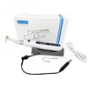
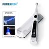
TECHNICAL PARAMETERS
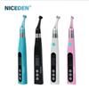
Brand name:NICEDEN
Battery: 1000mAh/3.7V
Input: 5V 1A
Output: 1.8W Max
Main Unit Weight: 78g
Gross Package Weight: 420g
Lens Material: Stainless Steel
Disinfection: The lens can be disinfected at a high temperature of 135°C
Package Dimensions: 15 x 9.3 x 4.5 cm
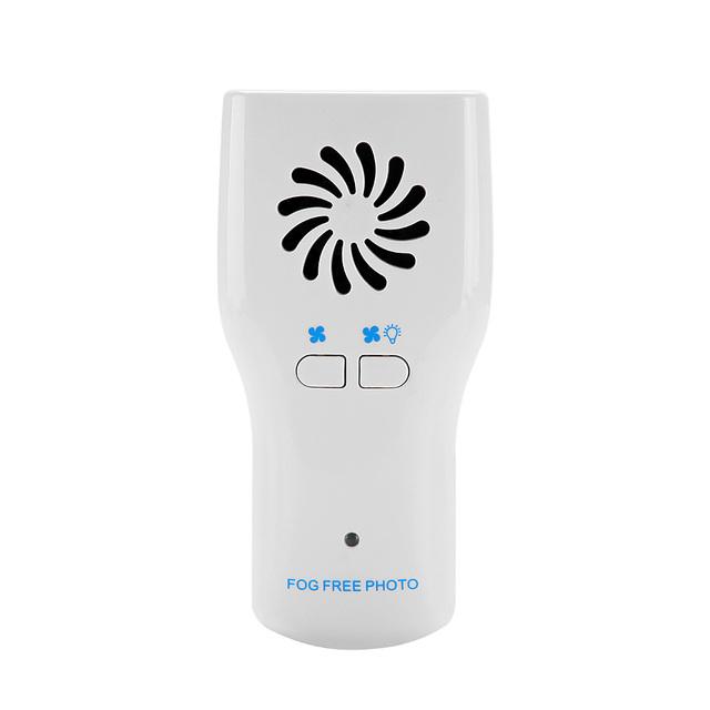
CHARGING INSTRUCTIONS
1.)When the indicator light flashes, brightness is insufficient, or the fan shuts down automatically, discontinue use of the device and recharge the battery immediately.
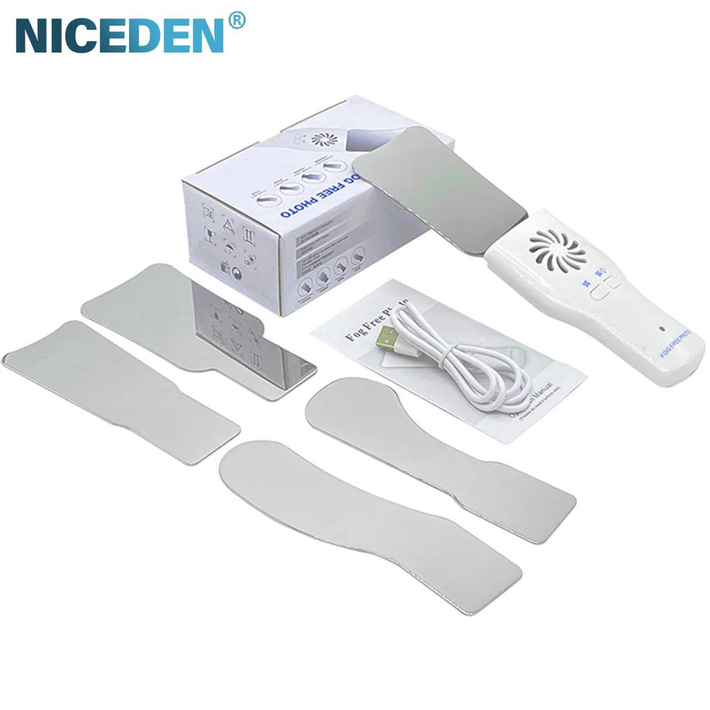
2.)Properly connect the AC adapter to the bottom socket of the device, then insert the AC USB adapter plug into a power outlet.
3.)A red indicator light illuminates during charging; the charging process is complete when the red indicator light switches to green.
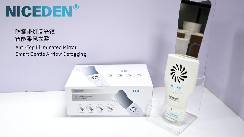
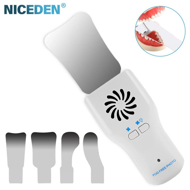
4.)Insert the mirror into the rear socket of the main unit, press the fan button, and a breeze will be emitted to defog the mirror surface. If brightness is insufficient, activate the lighting function (Note: Supplementary clarification based on device functionality as the original text contains a minor omission here).
5.)The device will automatically power off after 3 minutes of inactivity.
6.)After removing the mirror from the main unit, disinfect and clean the device in accordance with the user manual.
DISINFECTION, CLEANING, AND STERILIZATION
1.)The main unit and mirror must undergo cleaning, disinfection, and sterilization prior to each use to prevent contamination. This requirement applies to both the initial use and all subsequent uses.
2.)After each use, all components that have come into contact with infectious agents shall be cleaned using towels impregnated with a disinfectant-detergent solution (a bactericidal, fungicidal, and aldehyde-free solution). The use of chemical agents may potentially cause damage to the equipment.
3.)The mirror must be sterilized between treatments via autoclaving at 134℃.
4.)The main unit is not suitable for autoclaving.
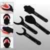
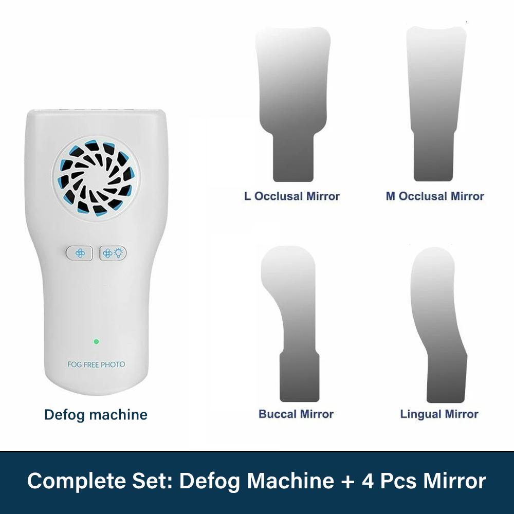
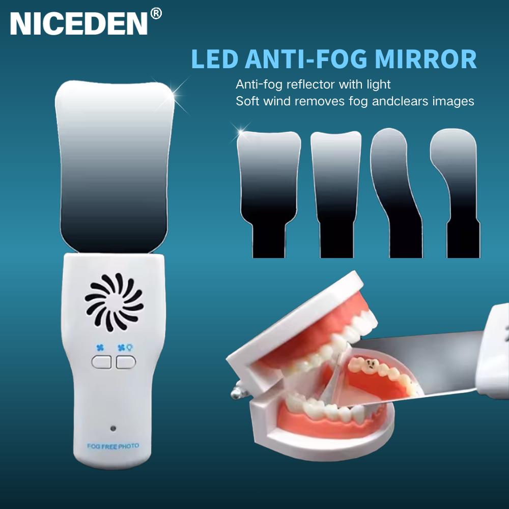
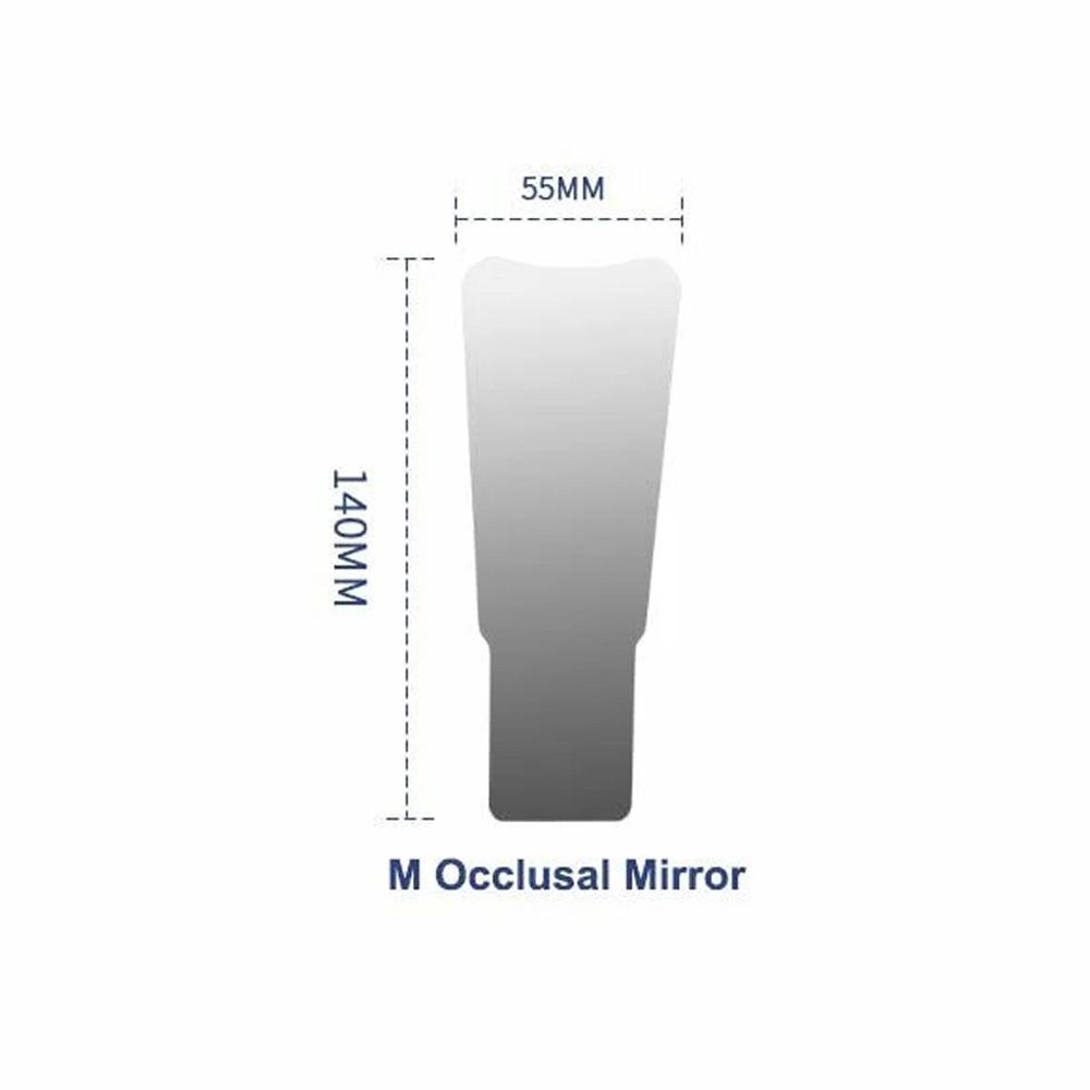
1.M - sized Occlusal Mirror
Routine examination: With a moderate size, it can be used for routine examinations in multiple areas of the oral cavity. It can clearly reflect the conditions of the occlusal surfaces (occlusal surfaces of the jaws) of the upper or lower teeth, facilitating dentists to check for caries, wear, cracks, and accumulation of dental calculus on the occlusal surfaces of molars, premolars, etc. For example, it can be used to check the degree of wear on the occlusal surfaces during the patient's chewing process and determine whether there is dentin hypersensitivity caused by excessive wear.
Treatment assistance: During treatments such as tooth filling and inlay restoration, it helps dentists observe whether the fit and shape of the restoration on the occlusal surface are appropriate, ensuring the normal occlusal function of the restored teeth. It can also assist in taking diagnostic images of the occlusal surfaces for comparison before and after treatment and for medical record keeping.
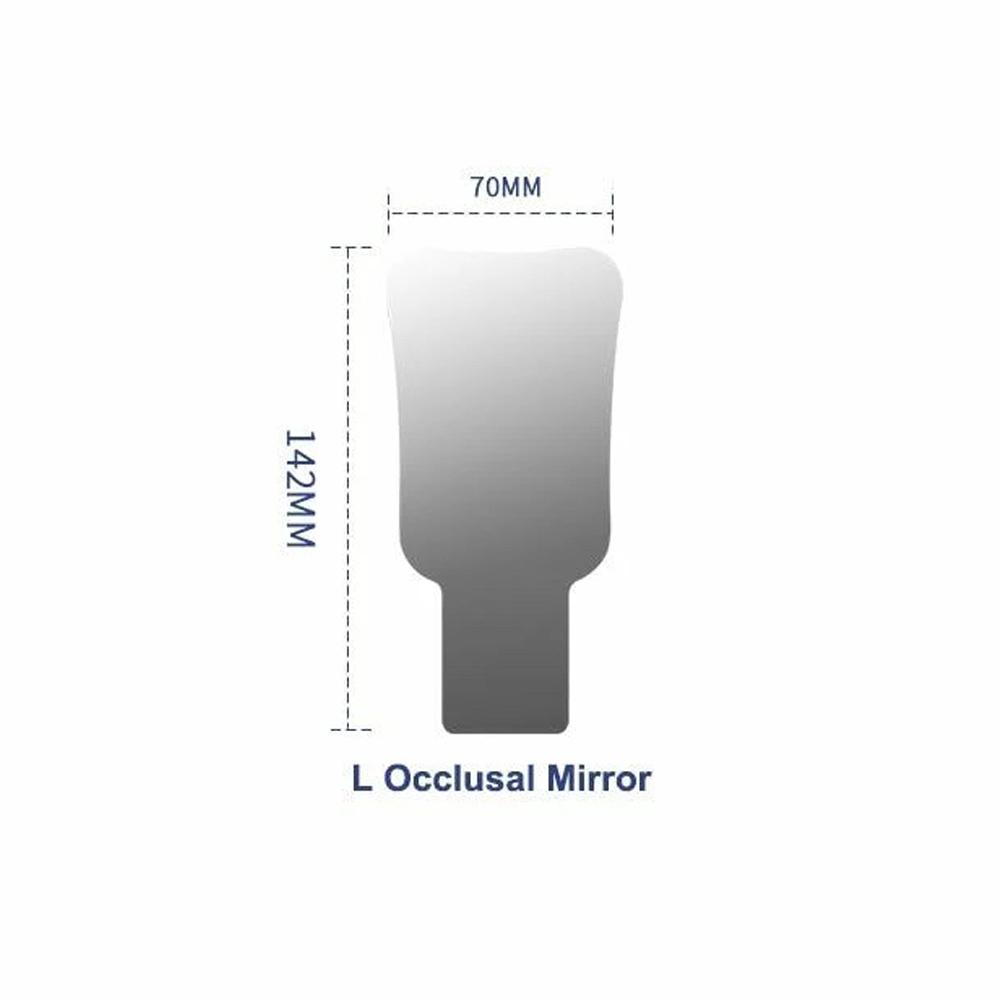
2.L - sized Occlusal Mirror
Wide - range observation: Due to its large size, it is suitable for observing the occlusal surface conditions of a relatively wide area in the oral cavity, such as the overall examination of the occlusal surfaces of all teeth. Especially during the initial oral examination and oral health screening, it can quickly check the general conditions of the occlusal surfaces of multiple teeth, determine whether there are common dental caries and dental calculus problems, and improve the examination efficiency.
Complex cases: For patients with disordered tooth arrangement and complex occlusal relationships (such as before orthodontic treatment for malocclusion), its large field of view can be used to observe the overall contact situation of the upper and lower teeth during occlusion and the occlusal interference points, providing references for the design of orthodontic treatment plans and occlusal adjustment. It is also beneficial for taking panoramic image data that includes the occlusal surfaces of more teeth.
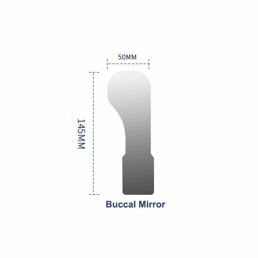
3.Buccal Mirror
Observation of buccal teeth and gums: It is mainly used to reflect the buccal surfaces (the surfaces of the teeth facing the inner side of the cheek) of the teeth and the corresponding buccal gums. Dentists can check for caries, attachment of dental calculus, abnormal tooth development (such as 斑块 on the buccal surfaces in dental fluorosis), etc. on the buccal surfaces, as well as the health problems such as color, swelling, bleeding, and recession of the buccal gums. For example, it can be used to check the buccal gum inflammation caused by the stimulation of poor restorations and evaluate the impact of gum health on the aesthetics and function of the teeth.
Assistance in aesthetic restoration: During cosmetic dental restoration (such as ceramic veneers and all - ceramic crown restorations), it helps dentists and patients observe the coordination of the color and shape of the restored buccal surfaces with the natural teeth and surrounding tissues. It can take comparison photos of the buccal surfaces before and after restoration for effect evaluation and doctor - patient communication, ensuring that the restoration meets the requirements of aesthetics and naturalness.
4.Tongue Depressor Mirror
Tongue depression and observation of the deep oral cavity: When in use, on the one hand, the mirror body is used to depress the tongue, avoiding the tongue from blocking the view of the deep oral cavity, and facilitating the examination of the conditions of the teeth (such as the distal surfaces of the lower molars), gums, and mucous membranes near the floor of the mouth. For example, it can be used to check whether there is caries on the distal surfaces of the last molars in the lower jaw, the state of the surrounding tissues during the eruption of wisdom teeth, and the lesions of the oral floor mucosa, etc. On the other hand, the mirror surface can reflect the images of the deep oral structures, assisting dentists to diagnose and record more clearly. For example, when taking diagnostic photos of the posterior part of the oral cavity, it can reduce the interference of the tongue and present clear images of the affected teeth and surrounding tissues.
Assistance in pharyngeal examination: In addition to the oral teeth and gums, during some simple pharyngeal examinations (in conjunction with the dental examination scenario), it can depress the tongue, allowing dentists to initially observe the general conditions of the posterior pharyngeal wall and tonsils, determine whether there are abnormalities such as redness, swelling, and secretions. Although it is not as comprehensive as professional pharyngeal examination instruments, it can be used as an auxiliary means for observing the oral - pharyngeal health in the dental diagnosis and treatment process.



 No-delivery refund